
 Front
Front
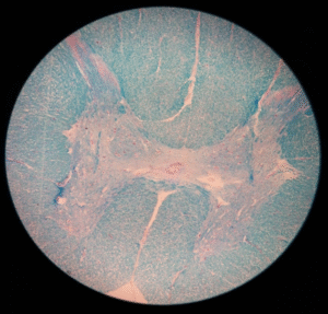
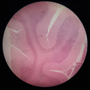
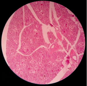
Thoracic Spinal cord
Identification Points:
1. Lateral gray horn.
2. White matter.
3. Gray commissure.
4. Central canal.
 Front
Front
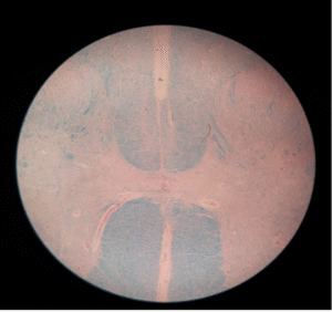
Cervical Spinal Cord
Identification Points:
1. Elongated posterior horn.
2. Later horn is absent.
3. Normal anterior horn.
4. Central canal.
 Front
Front
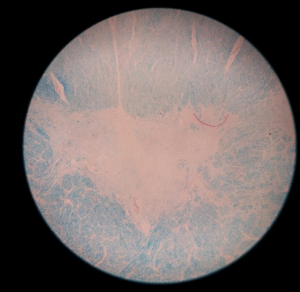
Lumbar Spinal Cord
Identification Points:
1. Gray matter is abundant compared to white matter.
2. Anterior and posterior horns are bulbous.
3. Gray commissure is thin.
4. Central canal.
 Front
Front
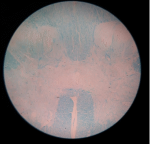
Sacral spinal cord
Identification Points:
1. Anterior and posterior horns are bulbous.
2. Lateral horn.
3. Thick gray commissure.
4. Central canal.
 Front
Front
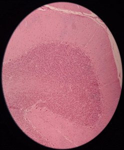
Cerebellar Cortex (4X)
Identification Points:
1. Outer Gray Matter.
2. Inner white matter.
3. Three layers: Granular layer, Perkinje layer and molecular layer.
 Front
Front
Cerebral Cortex (10X)
Identification Points:
1. Outer Gray Matter.
2. Inner white matter.
3. Six layers of gray matter.
 Front
Front
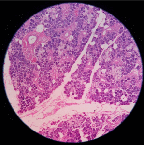
Submandibular Salivary Gland
Identification Points:
5. Mixed serous and mucous gland with predominance of serous alveoli.
6. Many serous demilunes.
7. Shorter and narrow intercalated ducts.
8. Numerous interlobular ducts.
Front
Sublingual Salivary Gland
Identification Points:
1. Mixed serous and mucous gland with predominance of mucous alveoli.
2. Few serous demilunes.
3. No intercalated ducts.
4. No capsule
Front
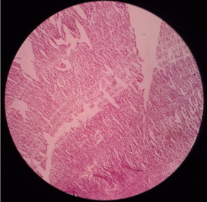
Parotid Salivary Gland
Identification Points:
1. Pure serious acini.
2. Striated ducts.
3. Interlobular ducts.
4. No serous demilunes.
Front
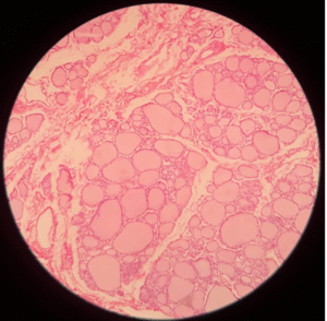
Thyroid Gland
Identification Points:
1. Characteristic follicles lined by simple cuboidal epithelium.
2. Very thin interfollicular stroma.
3. Numerous capillaries.
Front
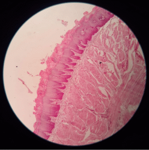
Tongue
Identification Points:
1. Lingual papillae of different size.
2. Lingual glands.
3. Skeletal muscles fiber running in all directions.
4. Stratified squamous non-keratinized epithelium
Front
Back